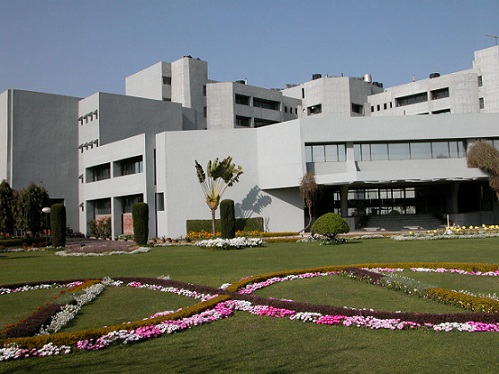
Dr Chayan Nandi (in the middle) with his team
Cell is the basic unit of life and it has several organelles that help it with many functions. Lysosome, present in animal cells, plays an important role in cellular processes and it is one of the tiniest cell organelles. Understanding the functional modalities of lysosome in live conditions is a challenging task even with the availability of high-end imaging techniques due to its very small size down to 50-500 nanometers (nm). Researchers from the Indian Institute of Technology Mandi (IIT Mandi) have designed gold nanoclusters as an efficient fluorescent nanoprobe for lysosome imaging.
The research team took up the image of the real size of lysosomes using a super-resolution microscopic imaging technique. The major challenge in super-resolution microscopic imaging is to get an efficient fluorescent probe, which at the same time with its intrinsic optical properties, can image the organelle to its real subnanometer size and also does not damage the cellular structure by posing any toxic effect.
The gold nanoclusters, because of its exciting photoluminescence properties, easy renal clearance from the body, and nontoxic nature, have tremendous biomedical applications. It has been used as a biomarker also. “Unfortunately, gold nanoclusters have never been explored for super-resolution microscopic imaging, we are the first to report its applicability as a fluorescent probe for single-molecule localization-based microscopic technique”, says Dr. Chayan Nandi, Associate Professor, School of Basic Sciences, IIT Mandi. “Because of its extremely small size, intrinsic fluorescence properties suitable for super-resolution microscopic imaging and the non-toxic nature, the gold nanocluster could be universally used for high-end optical imaging”, he added.
“ Gold nanoclusters have never been explored for super-resolution microscopic imaging, we are the first to report its applicability as a fluorescent probe for single-molecule localization-based microscopic technique”
In this work, the researchers have designed a bovine serum albumin protein conjugated red emissive gold nanoclusters as an efficient fluorescent nanoprobe for lysosome imaging. With this approach, the diameter of the lysosome is obtained as 59 nm, which is very close to the original diameter of the smallest lysosome in HeLa cells. HeLa is the oldest and most commonly used immortal cervical cancer cell line. They have been instrumental in studying human diseases, especially cancer.
The next step of this research is to make these gold nanoclusters as a universal probe for other multimodal and correlative bio imaging techniques. “Owing to its high electronic contrast, the developed nanoclusters, along with the use in super resolution optical microscopy, could also be used as a probe for transmission and scanning electron microscope in a correlative manner and thus will unveil a huge number of unresolved biological problems in live cell condition,” says Dr. Nandi.
The research team includes Dr Chayan Nandi, Dr Amit Jaiswal, Aditya Yadav, Navneet C. Verma, Chethana Rao and Pushpendra M. Mishra. The results of their work have been published in the Journal of Physical Chemistry Letters.
India Science Wire
ISW/JS/16/09/2020